Bone Cancer Prediction using machine learning
Bone Cancer Prediction
Price : 10000
Bone Cancer Prediction
Price : 10000
.svg)
ABSTRACT
Cancer is a dangerous disease, which is caused because of unregulated cell growth. After many researches, almost 100 different types of cancer have been detected in human body. Out of these, one of the most widely spread is bone cancer, which leads to death. The detection of bone cancer is very critical and which has no anticipation. Presently, most of the study is done by using data mining methods and the image processing techniques for medical image analysis process. Malignant and benign tumors of bone in the foot have traditionally been characterized as rare, or at least unusual. Bone cancer is considered to be the most dangerous and often the cause of early death around the globe. Therefore, early detection of the bone cancer has become needed to cure the patient. Medical imaging is playing an imperative function in analysis and healing of disease and locating tumours and finding of cancerous cells in premature phase. We Proposed a system to detect bone cancer accurately from MRI images using matlab and our proposed system will also classify images as cancerous or non cancerous.
INTRODUCTION
Bone is the supporting skeleton of body and is hollow. The outer part of bones is a arrangement of tough tissue called matrix against calcium salts are laid down. The hard out layer is made with cortical bone, it covers trabecular bone inside, outside of bone covered with periosteum. Some bones are hallow and space is called medullary cavity which contains the soft tissue called bone marrow. Endosteum is act as a tissue lining. At each end of the bone is a region of a softer shape of bone-like tissue called cartilage, it is softer than bone that is made of fibrous tissue matrix assorted with a gel-like stuff that does not enclose much calcium. Most bones get going out as cartilage. The body then put downs calcium onto the cartilage to form bone. After the bone formation, some cartilage may stay at the ends to act as a bolster between bones. This cartilage, along with ligaments and some other tissues join bones to form a joint. Bone itself is very stiff and muscular. Bone is able to hold up as much as 12,000 pounds per square inch. It takes as much as 1,200 to 1,800 pounds of pressure to break the thigh bone. The bone contains 2 kinds of cells. The osteoclast is the cell that forms new bone, and the osteoclast is the cell that softens old bone. Some bones the marrow is greasy tissue. The marrow in other bones is a concoction of fat cells and blood-forming cells. The blood-forming cells fabricate red blood cells, white blood cells, and blood platelets. Other cells in the marrow include plasma cells, fibroblasts, and reticuloendothelial cells. Cancer, which makes unfettered cell growth, will subdivide the cells and grow wildly, forming malevolent tumours, and assault nearby parts of the body.
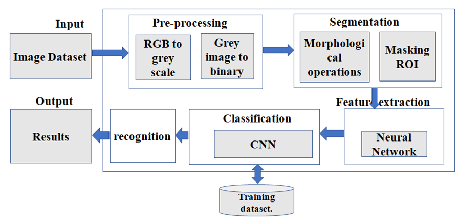
BLOCK DIAGRAM

SYSTEM REQUIREMENTS
A. Hardware Requirement:-
Ø System : Pentium IV 2.4 GHz.
Ø Hard Disk : 500 GB.
Ø Ram : 4 GB.
Ø Any desktop / Laptop system with above configuration or higher level.
B. Software Requirements:-
Ø Operating system : Windows XP / 7
Ø MATLAB 2015 and above with following tool boxes
ü Image Processing
ü Machine Learning
Conclusion
Bone cancer is one kind of dangerous diseases, so it is necessary to detect cancer in its early stages. But the detection of bone cancer is the most difficult task. From the literature review, many techniques are used for the detection of bone cancer but they have some limitations. In our proposed method pursue approaches in which the first step is preprocessing, edge detection, morphological operation, segmentation and then feature extraction. The proposed system aims detects the bone cancer from MRI scan images. The system achieves its desired expectation at the end of the system. The extracted features from the image contain some specific information to understand the details of the image. The main purpose of extracting the features is to reduce the process complication and also to isolate various desired shape of the image. The accuracy of the classification stage depends on extracted features.